
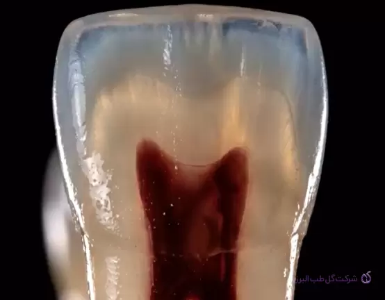
Successful root canal treatment is not limited to understanding instruments and canal preparation techniques. A thorough understanding of root canal system anatomy is one of the most important factors in accurate diagnosis, cleaning, shaping, and ultimately successful obturation.
A root canal does not always follow a simple, straight path from the pulp chamber to the apex. Depending on the type of tooth and even the individual roots of a multirooted tooth, canals may exhibit branching, merging, separate pathways, and multiple apical foramina. Therefore, identifying the canal configuration before treatment can help prevent residual pulp tissue, persistent microbial contamination, and eventual treatment failure.
One practical classification used to describe canal configurations is the Weine classification, which categorizes canal patterns according to the number of canals and their pathways toward the root apex.
Why Is Identifying the Canal Type Important?
Canal classification is not merely theoretical; it can directly influence clinical treatment decisions.
Understanding canal anatomy helps clinicians make more informed decisions throughout different stages of treatment, including:
-
Canal identification and location
-
Determination of the canal pathway and working length
-
Cleaning and mechanical preparation
-
Root canal irrigation
-
Identification and management of accessory canals and branches
-
Selection of an appropriate obturation technique
-
Radiographic evaluation of treatment outcomes
The greater the anatomical complexity, the higher the risk of leaving portions of the root canal system unidentified or inadequately cleaned.
Canal Classification According to Weine
In the classic Weine classification, canal configurations are described according to the number of canals at the beginning of the root canal pathway and the number of apical foramina.
The four main configurations are as follows.
Type I — 1-1
One canal, one foramen
In this configuration, a single canal originates from the floor of the pulp chamber and continues toward the apical region without dividing or merging with another canal. It ultimately exits through a single apical foramen.
This is one of the simplest and most common anatomical configurations in single-canal roots.
Pathway:
Pulp chamber → One canal → One apical foramen
Type II — 2-1
Two canals, one foramen
In this configuration, two separate canals originate from the pulp chamber but merge along their course and exit through a common apical foramen.
This pattern is clinically important because two canals may appear completely separate at the beginning of treatment but communicate and merge in the deeper portions of the root.
Pathway:
Two separate canals → Apical merging → One foramen
Therefore, observing a single apical foramen on a radiograph does not necessarily mean that only one canal exists throughout the entire root.
Type III — 2-2
Two canals, two foramina
In this configuration, two separate canals originate from the pulp chamber and remain independent throughout their course. Each canal terminates through its own apical foramen.
Pathway:
Two separate canals → Two independent pathways → Two apical foramina
This configuration has particular clinical significance because both canals must be independently identified, prepared, irrigated, and obturated.
Type IV — 1-2
One canal, two foramina
In this configuration, a single canal originates from the pulp chamber but divides into two separate pathways in the apical portion of the root, eventually exiting through two distinct apical foramina.
Pathway:
One canal → Apical division → Two foramina
This configuration can make diagnosis and treatment challenging because the canal may appear as a single pathway in the coronal portion, while the main division occurs deeper within the root.
An Important Distinction Between Type II and Type III
One of the key aspects of the Weine classification is the difference between Type II and Type III.
Both configurations begin with two canals, but the main difference lies in what happens to these canals along their course.
Type II (2-1): The two canals merge and exit through a single apical foramen.
Type III (2-2): The two canals remain independent until the root apex and terminate through separate foramina.
Therefore, the number of canals at the beginning of the pathway alone is insufficient to distinguish these two configurations. Their course toward the apical region must also be evaluated.
In contrast, Type IV is the only one of these four main Weine configurations that begins with one canal and terminates through two apical foramina.
Important Note: The Weine Classification Applies to Individual Roots
A practical consideration when using this classification is that the canal type should be described for each root individually, rather than only for the tooth as a whole.
This is particularly important in multirooted teeth, where different roots of the same tooth may exhibit different anatomical configurations.
Therefore, when describing the anatomy of a multirooted tooth, the canal configuration of each root should be assessed separately.
Clinical Challenges in Complex Root Canal Anatomy
As the root canal system becomes more complex, the risk of errors during diagnosis, canal location, preparation, and obturation increases.
In some cases, the canal configuration may not be clearly visible on conventional radiographs. Two canals may also be present within the same root but appear as a single pathway because their radiographic images overlap.
For this reason, the suspected canal configuration should always be evaluated alongside the clinical examination and careful interpretation of radiographic images.
Type IV Canals in Mandibular Premolars: A Significant Challenge
One anatomical configuration that can complicate root canal treatment is Type IV (1-2) in mandibular premolars.
In these cases, a single canal is visible in the coronal portion but divides into two canals in the deeper portions of the root, eventually terminating through two separate apical foramina.
The main challenge is that the division may not be clearly visible on conventional radiographs, and the second canal may remain undetected during the initial stages of treatment.
Depending on the patient's condition and the complexity of the case, more advanced diagnostic and treatment approaches may be beneficial.
These may include:
-
Limited-field-of-view CBCT (Limited FOV CBCT) in selected cases
-
Magnification and the use of a dental operating microscope
-
Adequate illumination and careful examination of the pulp chamber floor
-
Effective active irrigation techniques
-
Selection of an appropriate obturation technique, including Warm Vertical Condensation when indicated
The goal is to improve the identification of hidden canal pathways, enhance cleaning of the root canal system, and achieve an appropriate seal throughout the prepared canal space.
Identifying Type II and Type III Canals in Mandibular Molars
Another important clinical situation involves the mesial roots of mandibular molars, where two mesial canals may exhibit different configurations, including Type II or Type III.
This distinction can be particularly relevant in selected retreatment cases, cases involving instrument fracture or separation, and situations in which previous treatment has been complicated.
In these circumstances, relying on a single radiographic image may not provide sufficient information.
Radiography at Different Angulations
Taking radiographs at different horizontal angulations can help distinguish overlapping canal pathways.
Changing the horizontal angle of the X-ray beam may separate canals that appear superimposed in a straight-on projection.
In many cases, this simple technique can provide valuable information about the presence of a second canal and its possible course.
Using Paper Points or Gutta-Percha Cones
In selected cases, placing a paper point, gutta-percha cone, or hand file within the canal and evaluating its position radiographically can help clarify the canal pathway and identify changes in its configuration.
When interpreted alongside other clinical and radiographic findings, this approach may provide additional information about canal separation, merging, or branching.
The Role of Magnification and Illumination in Locating Additional Canals
One of the key factors in identifying complex anatomy is creating optimal conditions for visualizing the treatment field.
Magnification, adequate illumination, and careful examination of the pulp chamber floor can increase the likelihood of locating additional canals.
A dental operating microscope can be particularly valuable when canal orifices are small, calcified, or located in atypical positions.
In complex endodontic treatment, success depends not only on having the right instruments but also on the ability to visualize and interpret the anatomy accurately.
When Can CBCT Be Helpful?
CBCT can provide valuable information when root canal anatomy cannot be adequately assessed using two-dimensional imaging.
It may be particularly useful in selected cases involving:
-
Suspected additional canals
-
Complex root anatomy
-
Canal branching
-
Endodontic retreatment
-
Suspected untreated canals
-
Unusual canal configurations
However, CBCT should be prescribed according to diagnostic necessity and the principle of justification. When indicated, a limited field of view may be more appropriate for detailed assessment of the region of interest.
From Diagnosis to Obturation: Why Does Canal Type Matter?
Identifying the canal type is important not only for locating canals but also for improving the quality of subsequent treatment stages.
In a complex canal system, an unidentified pathway or branch may remain outside the effective reach of cleaning and irrigation procedures.
The obturation technique should also be selected according to the canal anatomy.
Oval canals, apical branches, merging canals, and other complex configurations may require particular attention to achieving a three-dimensional seal.
In selected complex cases, techniques such as Warm Vertical Condensation may help improve gutta-percha adaptation to the prepared canal space. However, the appropriate obturation technique should always be selected according to the individual case and clinical judgment.
Conclusion
Root canal system anatomy is far more diverse than what may be apparent on a conventional radiograph.
The Weine classification provides a practical framework for understanding several major anatomical configurations by describing the relationship between the number of canals and the number of apical foramina.
|
Type |
Canal configuration |
Description |
|---|---|---|
|
Type I |
1-1 |
One canal, one foramen |
|
Type II |
2-1 |
Two canals merging into one canal and one foramen |
|
Type III |
2-2 |
Two independent canals and two foramina |
|
Type IV |
1-2 |
One canal dividing into two canals and two foramina |
Ultimately, understanding these configurations becomes most valuable when combined with clinical examination, appropriately angulated radiographs, magnification, adequate illumination, and CBCT in selected cases.
In endodontics, what remains unseen can be just as important as what is visible. Accurate identification of root canal anatomy is the first step toward effective cleaning, appropriate shaping, and reliable obturation of the root canal system.